Cardiology tests available
Cardiology tests available
At Complete Cardiology we offer the basic cardiac tests in outpatient settings at The Bridge Clinic in Maidenhead and Princess Margaret Hospital in Windsor. These include the following:
More complex tests are referred to our consultant partners in more specialist hospitals, such as the John Radcliffe Hospital in Oxford and Harefield Hospital.
Electrocardiogram (ECG)
This simple, painless test takes a few minutes to perform. It turns the normal electrical activity of the heart into a tracing and this can reveal a number of different cardiac conditions which affect the structure and rhythm of the heart. As it is only a single brief snapshot of the heartbeat at rest a normal ECG does not necessarily exclude important disease of the heart but it is a good start!
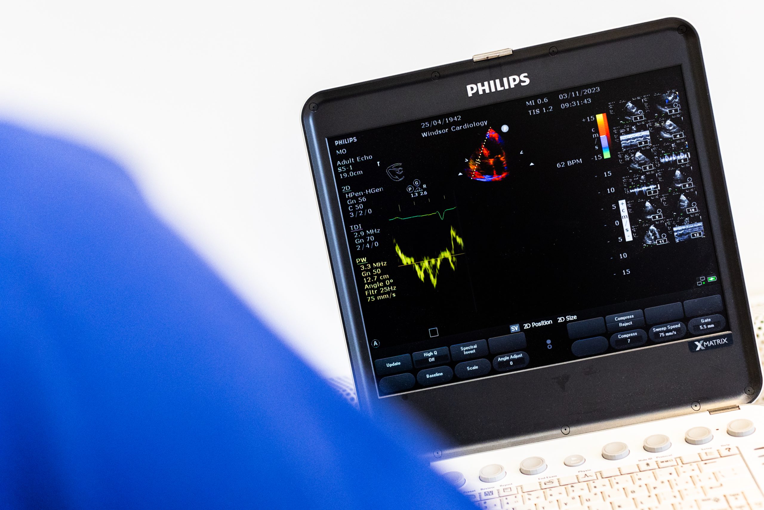
Echocardiogram (Echo)
This completely safe and painless test takes up to 30 minutes to perform and revolutionised the assessment of cardiac disease when it was developed in the 1970s. It uses exactly the same technology as used in ultrasound to look at babies developing in the womb and gives high definition moving images of the heart. This allows us to assess all aspects of heart function including size and thickness of the walls and chambers of the heart, the hearts pumping function and complete assessment of the valves of the heart.
Our own state-of-the-art dedicated cardiac echocardiography machine will be available during every clinic session.
Blood Tests
Blood testing is a fundamental way of checking for disease and establishing the chances or risk of developing a heart problem in the future.
We have access to the full range of blood tests and the results will be available promptly – sometimes even on the day they are taken.
24 Hour Blood Pressure Recording
High blood pressure levels (often called hypertension) are one of the major causes or risk factors for the development of heart disease and stroke. It is normal for blood pressure readings to vary throughout the day but it is well known that some patients have very high readings when they are taken in a medical setting (white coat effect) but overall their ‘true’ blood pressure is satisfactory. If this situation is suspected we may recommend you wear a special machine which takes and records your blood pressure hourly throughout the day and night. This gives us a feel for your true blood pressure. The cardiology tests may also be used to check that satisfactory control is being achieved in patients taking tablets for high blood pressure.
ECG monitors (‘Holter’ recording)
Symptoms of palpitations or awareness of the heartbeat are extremely common but usually only occur occasionally. A simple resting ECG will often not ‘catch’ the rhythm of the heart whilst the patient has symptoms. In this situation we will often suggest the fitting of a 24- or 48-hour ECG recorder. This lightweight device is made of thin silicon and is attached to the chest with two electrodes. It records the heartbeat continuously for 24 or 48 hours and provides valuable information about the basic rhythm of the heart as well as the rhythm at the time of symptoms.
Sometimes palpitation symptoms are very elusive and typically disappear for the 24/48 hours of recording. In this situation a longer period of recording (‘7 or 14-day event recorder’) may be recommended.
Coronary CT scanning and EBCT (Electron-beam CT scan)
This is a scan to look for calcium build up in the arteries of the heart (coronary calcification) which is a very early sign of furred and narrowed arteries. This usually occurs long before any symptoms of a heart problem develop and is therefore used as a screening test in people who may be at risk of heart disease. This is called calcium scoring but has largely been superseded by CT coronary angiography which gives detailed information of the arteries of the heart including chalk and fat deposits and the degree of narrowing within the heart arteries.
Thallium Scan (Perfusion scan)
This is a specialised scan to assess the blood flow to the heart during exercise. It is similar to an exercise ECG test but may give more precise information and can be used in patients unable to exercise on a treadmill. When exercising, a tiny amount of a special radioactive substance is injected and a scan of the heart taken. Areas of the heart with poor blood supply are highlighted on the scan.
If this test is not normal your cardiologist may well suggest you have an angiogram.
Exercise Tolerance Test (Exercise Test)
This is used to be a key test to assess angina and is still used by some medical screening companies to ‘screen’ patients with no symptoms at all for early signs of coronary heart disease. A continuous ECG is taken whilst you undergo a graded standardised exercise on a treadmill. The test starts at a leisurely pace and gradually the speed and gradient increase every three minutes.
Patients with angina will typically experience symptoms and this is associated with characteristic changes to the shape of the ECG.
Specialised Echo
Echocardiogram and ultrasound technology has developed very rapidly and in certain situations even more specialist forms of echo scanning of the heart is required.
Trans-Oesophageal Echo (T.O.E.)
Usually, a standard echocardiogram is detailed enough to give all the information about the heart we require. Sometimes however, particularly in patients with a problem with one of the heart valves, we need to get really detailed pictures of the heart and this can be done by passing a narrow flexible tube down your throat and gullet. The tube has a tiny echo sensor built into the tip and this generates extremely detailed pictures of the heart with no interference from the lungs and ribs. This procedure is often also performed during heart valve surgery to help guide the surgeon. This test is extremely safe and you will usually only need to spend a few hours in the hospital and go home on the same day. It requires a brief general anaesthetic.
Stress Echo
Even in patients with severe narrowing of the hearts arteries (coronary artery disease) the echocardiogram at rest may be completely normal. In this situation however under the stress of exercise the function of the heart can become abnormal in areas of the heart where there is a problem with blood flow. Stress echo uses this fact to help us diagnose and treat coronary artery disease and may be used as an alternative to an exercise test or perfusion (thallium) scan. Following taking a detailed resting echocardiogram the heart is ‘stressed’ either by exercising you or with a special medicine injected into a small vein. The echocardiogram is then repeated and the images compared.
This test is extremely safe and you will usually only need to spend an hour or two in the hospital and go home on the same day.
Tilt Testing
This test is used to investigate dizziness and fainting. The patient is placed on a specially designed bed which is then tilted to 60 degrees with the head up. The patient’s pulse and blood pressure are closely monitored. It has been found that, depending on the cause, this test will provoke a typical episode of fainting and thereby allow us to see the cause of the problem and plan treatment.
Angiogram (also coronary angiogram, cardiac catheterisation)
This is a special x-ray test to take pictures of the arteries of the heart. It is a very safe test and is performed under local anaesthetic. A tiny tube is passed to the heart via an artery in either the leg or wrist and moving x-ray pictures recorded as a special dye is injected into the arteries. This gives us a road map of the arteries and shows up any blockages or narrowing. During this test the valves, pumping function and oxygen levels in the heart can also be assessed.